Differences between digital pathology and traditional pathology
Digital pathology and traditional pathology are two different approaches to analyzing tissue samples for the diagnosis of disease. Both methods involve the examination of tissue samples under a microscope, but there are key differences in the way that samples are prepared, analyzed, and stored. Traditional pathology, also known as “analog pathology,” involves the processing of tissue samples and their preparation as glass slides. Once prepared, the slides are examined under a microscope by a pathologist, who will make observations and annotations by hand. This method has been used for over a century and is still widely used in pathology labs around the world. Digital pathology, on the other hand, involves the conversion of tissue samples into digital images using a digital slide scanner. The images can then be viewed, annotated, and analyzed on a computer using image analysis software. This allows for faster, more accurate, and more efficient diagnoses, as well as new opportunities for research. Additionally, digital pathology enables the easy sharing of images and information, which improves collaboration and increases the speed of diagnoses.
Another important difference between digital pathology and traditional pathology is the way images are stored. In traditional pathology, slides are typically stored physically in a slide library, which can make it difficult to access and share images. Digital pathology, however, allows for the electronic storage and retrieval of images, which eliminates the need for physical storage and allows for easy access to images. In summary, digital pathology and traditional pathology are two different approaches to analyzing tissue samples that have their own advantages and disadvantages. Digital pathology is a more recent technology that offers faster, more accurate, and more efficient diagnoses, as well as new opportunities for research, while traditional pathology is widely used and offers a more established workflow.
Overview of traditional pathology workflow, including tissue processing and slide preparation
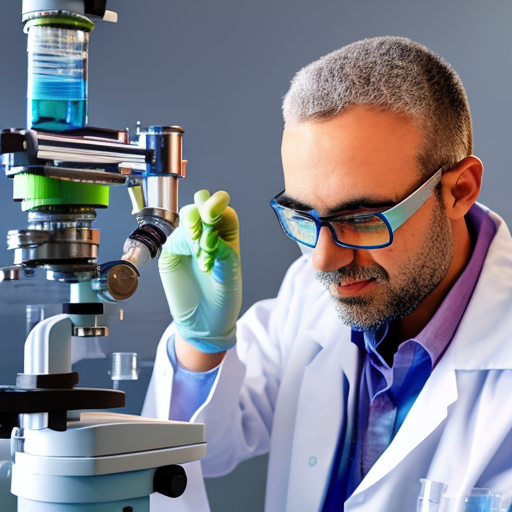
Traditional pathology workflow begins with the collection of tissue samples, which can be obtained through biopsies or during surgical procedures. These samples are then processed in a laboratory setting, where they are fixed, embedded, and cut into thin sections. The sections are then mounted onto glass slides, which are labeled and stored for examination by a pathologist.
The tissue processing stage is crucial for obtaining high-quality sections that can be easily examined under a microscope. It involves the fixation of the tissue samples, which preserves the tissue structure and prevents further decay. The samples are then embedded in paraffin, which allows for the cutting of thin sections that can be mounted onto glass slides. The sections are stained with dyes that highlight specific structures or cellular components, making it easier for the pathologist to observe and analyze the tissue. Once the slides are prepared, they are examined by a pathologist, who will make observations and annotations by hand, using a microscope. The pathologist will then make a diagnosis based on their observations and the results of any additional testing that may have been performed on the tissue sample.
Overview of digital pathology workflow, including digital slide scanning and image analysis
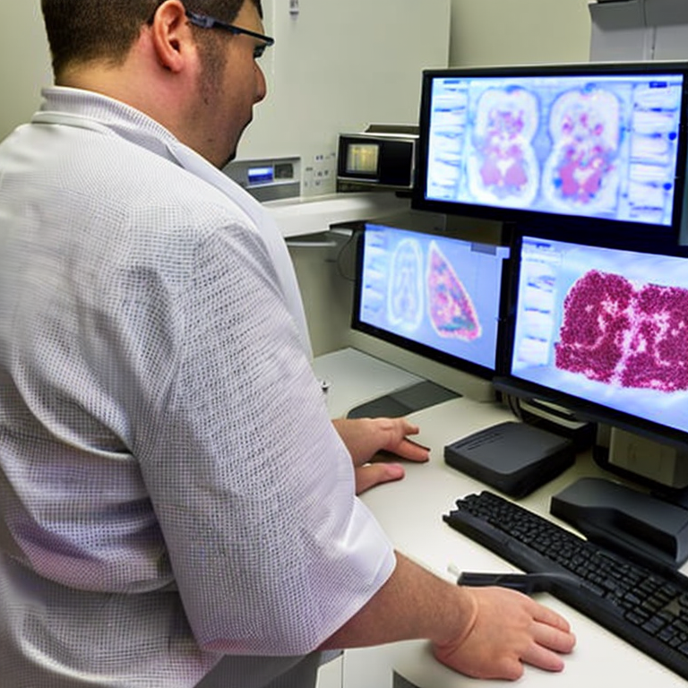
Digital pathology workflow begins with the collection of tissue samples, which can be obtained through biopsies or during surgical procedures. These samples are then processed in a laboratory setting, where they are fixed, embedded, and cut into thin sections. The sections are then scanned using a digital slide scanner, which converts the tissue sections into high-resolution digital images.
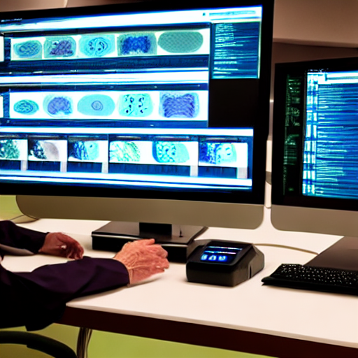
Once the tissue samples have been scanned, the digital images can be viewed, annotated, and analyzed on a computer using image analysis software. This allows for faster, more accurate, and more efficient diagnoses, as well as new opportunities for research. Additionally, digital pathology enables the easy sharing of images and information, which improves collaboration and increases the speed of diagnoses. The digital images can be stored electronically, which eliminates the need for physical storage and allows for easy access to images. The pathologist can access these images remotely and make observations and annotations digitally, using a computer. The pathologist will then make a diagnosis based on their observations and the results of any additional testing that may have been performed on the tissue sample. The software also allows for the use of artificial intelligence and machine learning to improve the accuracy and speed of diagnoses.
Comparison of the time and efficiency of traditional vs. digital pathology workflow
One of the main advantages of digital pathology over traditional pathology is the increased speed and efficiency of the workflow. Traditional pathology relies on manual examination of glass slides under a microscope, which can be time-consuming and subject to human error. In contrast, digital pathology allows for the rapid examination of digital images using image analysis software, which can speed up the diagnostic process.
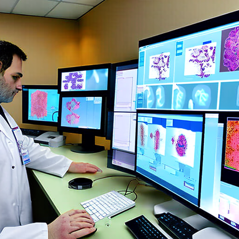
In addition, digital pathology enables the simultaneous review of multiple images by multiple pathologists, which can further increase the speed of diagnoses. This can be particularly useful in remote or underserved areas, where access to a pathologist may be limited. Digital pathology also allows for the collection and analysis of large amounts of data, which can be used for tasks such as drug development, identifying new biomarkers, and understanding disease mechanisms. This can be a significant advantage over traditional pathology, which often relies on small sample sizes and limited data.
On the other hand, traditional pathology workflow has its own advantages. For instance, it allows for the examination of samples under a microscope, which can be important in certain cases, such as the study of fine structures or the detection of certain diseases. Additionally, traditional pathology has been used for over a century and is well-established, which can be an advantage in certain cases.
Advantages and disadvantages of traditional vs. digital pathology workflow
Traditional pathology has some advantages over digital pathology. For example, it allows for the examination of samples under a microscope, which can be important in certain cases, such as the study of fine structures or the detection of certain diseases. Additionally, traditional pathology has been used for over a century and is well-established, which can be an advantage in certain cases. It also allows for the use of specialized stains and techniques that may not be available in digital pathology.
However, digital pathology also has its own advantages. One of the main advantages is the increased speed and efficiency of the workflow. Digital pathology allows for the rapid examination of digital images using image analysis software, which can speed up the diagnostic process. Additionally, digital pathology enables the simultaneous review of multiple images by multiple pathologists, which can further increase the speed of diagnoses. This can be particularly useful in remote or underserved areas, where access to a pathologist may be limited. Digital pathology also allows for the collection and analysis of large amounts of data, which can be used for tasks such as drug development, identifying new biomarkers, and understanding disease mechanisms.
On the other hand, digital pathology workflow also has some disadvantages. One of the main challenges of digital pathology is the need for high-resolution images, which can be challenging to obtain. Additionally, image analysis software can be complex and difficult to use, which can limit its application. There are also concerns about the accuracy and reliability of digital diagnoses and the potential for data breaches.
The impact of digital pathology on pathology workflow and efficiency
The introduction of digital pathology has had a significant impact on the workflow and efficiency of pathology practice. One of the main benefits of digital pathology is the ability to share images and information easily, which can improve collaboration and increase the speed of diagnoses. This is especially important in remote or underserved areas, where access to a pathologist may be limited. Digital pathology also allows for the collection and analysis of large amounts of data, which can be used for tasks such as drug development, identifying new biomarkers, and understanding disease mechanisms. This can be a significant advantage over traditional pathology, which often relies on small sample sizes and limited data.
Digital pathology also improves the efficiency and accuracy of pathology workflows. For example, digital images can be reviewed remotely, which allows for the rapid exchange of information and expertise. Digital images can also be stored and accessed electronically, which eliminates the need for physical storage and allows for easy retrieval. Additionally, the use of artificial intelligence and machine learning in digital pathology can improve the accuracy and speed of diagnoses, which can ultimately lead to better patient outcomes.
Overall, the introduction of digital pathology has had a positive impact on pathology workflow and efficiency. It has improved collaboration and speed of diagnoses, and has also opened new opportunities for research. Additionally, digital pathology has made the workflow more efficient and accurate, which ultimately leads to better patient outcomes.