
Our goal is to integrate artificial intelligence in developing infrastructure and work-flows for digital diagnostic pathology, facilitating transformational changes in the detection and treatment of disease, improving patient safety, and thus promoting sustainability of the Ontario health system.
A university-industry-government partnership is developing new artificial intelligence/machine learning tools for the next generation of digital pathology scanners. The project aims to revolutionize pathology infrastructure in Ontario hospitals, facilitating transformational changes in the detection and treatment of disease, improving patient safety, and promoting sustainability of the Ontario health system.
>The project has three main research objectives:
The project's thorough validation will take place at St. Michael's Hospital in Toronto and Toronto General Hospital/Research Institute (UHN). Commercial and social benefits are expected to accrue from the scanning systems and software products developed. Huron Digital Pathology will provide the means for developing, commercializing, and applying the research outcomes.
This government-funded project on transforming digital pathology with AI is led by a team of scientists and physicians from the University of Toronto, industry partner Huron Digital Pathology, University of Waterloo, Queens University, and government partners. The project will also recruit students and associates to work on the project. The team's expertise in pathology, machine learning, and computer engineering will allow them to create a comprehensive digital pathology database, develop efficient neural networks, and create an AI-powered diagnosing system. The validation of the project will take place at St. Michael's Hospital in Toronto and Toronto General Hospital/Research Institute (UHN). The commercial and social benefits from this project will be enormous, as it will provide accurate, faster, and cost-effective healthcare decisions regarding diagnosis and treatment at Ontario hospitals.
Principle Investigator
Konstantinos (Kostas) N. Plataniotis received his B. Eng. degree in Computer Engineering from University of Patras, Greece and his M.S. and Ph.D. degrees in Electrical Engineering from Florida Institute of Technology Melbourne, Florida. Dr. Plataniotis is currently a Professor with The Edward S. Rogers Sr. Department of Electrical and Computer Engineering at the University of Toronto in Toronto, Ontario, Canada, where he directs the Multimedia Laboratory and the University of Toronto-Huawei Mobile AI Laboratory. He holds the Bell Canada Endowed Chair in Multimedia since 2014. His research interests are primarily in the areas of image/signal processing, machine learning and adaptive learning systems, visual data analysis, multimedia and knowledge media, and affective computing.
Dr. Plataniotis is a Fellow of IEEE, Fellow of the Engineering Institute of Canada, Fellow of the Canadian Academy of Engineering / L’ Academie Canadienne Du Genie, and a registered professional engineer in Ontario. He has served as the Editor-in-Chief of the IEEE Signal Processing Letters. He was the Technical Co-Chair of the IEEE 2013 International Conference in Acoustics, Speech and Signal Processing, and he served as the inaugural IEEE Signal Processing Society Vice President for Membership (2014 -2016) and General Co-Chair for the 2017 IEEE GLOBALSIP. He served as the 2018 IEEE International Conference on Image Processing (ICIP 2018), and as General Co-Chair of the 2021 International Conference on Acoustics, Speech and Signal Processing (ICASSP21). He will be the General Chair of the 2027 IEEE International Conference on Acoustics, Speech and Signal Processing (ICASSP2027).
Co-PI (VP-Research Technology)
Savvas Damaskinos is the VP-Research Technology at Huron Digital Pathology. He joined the company (originally known as Biomedical Photometrics Inc.) in 1998 as project leader. He led the development and manufacturing of micro array readers, tissue imaging systems, and a MacroScope/Microscope combination system for general confocal imaging applications. He has been involved in the development of imaging systems for more than 20 years and is the author of several patents and more than 30 publications on confocal microscopy imaging techniques and applications.
Savvas Damaskinos received his Ph.D. in Physics from the University of Waterloo in 1985 and was the first recipient of the GWP Pearson medal for graduate work in Physics. He spent three years at the Institute of Energy Conversion in Delaware, a centre of excellence for photovoltaics, r eturning to the Department of Physics at the University of Waterloo as a Research Associate Professor in 1988. Research activities at the Physics Department resulted in several patents in the area of confocal scanning laser imaging technology.
Co-PI (ECE Professor)
Andreas Moshovos along with his students has been answering the question “what is the best possible digital computation structure to solve problem X or to run application Y?” where “best” is a characteristic (or combination thereof) such as power, cost, complexity, etc. Much of his work has been on high-performance processor and memory system design and it has influenced commercial designs. Andreas Moshovos has received the Ptyhio and a Master’s in Computer Science from the University of Crete in 1990 and 1992 and the PhD degree in Computer Sciences from the University of Wisconsin-Madison in 1998. He has taught Computer Design at Northwestern University, USA, (Assistant Professor 1998-2000), the Ecole Polytechnique de Laussane, Switzerland, (Invited Professor 2011) and since 2000 at the Electrical and Computer Engineering Department of the University of Toronto where he now is a professor.
Andreas Moshovos has served as the Program Chair for the ACM/IEEE International Symposium on Microarchitecture in 2011 and on numerous technical program committees in the area of Computer Architecture. He is an Associate Editor for the IEEE Computer Architecture Letters and the Elsevier Journal on Parallel and Distributed Computing.
Co-PI (ECE Professor)
Zhou Wang received the Ph.D. degree from The University of Texas at Austin in 2001. He is currently a Canada Research Chair and Professor in the Department of Electrical and Computer Engineering, University of Waterloo, Canada. His research interests include image and video processing and coding; visual quality assessment and optimization; computational vision and pattern analysis; multimedia communications; and biomedical signal processing.
Prof. Wang serves as a member of IEEE Image, Video and Multidimensional Signal Processing Technical Committee (2020-2022) and IEEE Multimedia Signal Processing Technical Committee (2013-2015), a Senior Editor of IEEE Journal of Selected Topics in Signal Processing (2022-present), a Senior Area Editor of IEEE Transactions on Image Processing (2015-2019), and an Associate Editor of IEEE Transactions on Circuits and Systems for Video Technology (2016-2018), IEEE Transactions on Image Processing (2009-2014), and IEEE Signal Processing Letters (2006-2010), among other journals. He was elected a Fellow of Royal Society of Canada: Academy of Science in 2018, a Fellow of Canadian Academy of Engineering in 2016, and a Fellow of IEEE in 2014. He is a recipient of 2021 Excellence of Graduate Supervision Award at University of Waterloo Faculty of Engineering, 2021 Technology Emmy Award, 2016 IEEE Signal Processing Society Sustained Impact Paper Award, 2015 Primetime Engineering Emmy Award, 2014 NSERC E.W.R. Steacie Memorial Fellowship Award, 2013 IEEE Signal Processing Magazine Best Paper Award, and 2009 IEEE Signal Processing Society Best Paper Award.
Co-PI (Anatomical Pathology)
Dr. Rowsell is an Associate Professor of Laboratory Medicine and Pathobiology at the University of Toronto and Division Head of Pathology at Unity Health Toronto. His academic interests include gastrointestinal pathology, quality assurance, and application of new technologies in Pathology. He has held several leadership roles in quality assurance, including Chair of the Pathology Scientific Committee of the Institute for Quality Management in Healthcare (IQMH), and was a member of the Advisory Panel for the Ontario provincial Quality Management Program (QMP).
Dr. Rowsell is also strongly engaged in MD education, and was awarded the Kalman Kovacs Undergraduate Medical Education Award (2018), the MD Program Teaching Award for Excellence (2018, 2020, 2021), and the Norman Rosenblum Award for Mentorship in the MD/PhD Program (2019). He has a strong history of collaboration in interdisciplinary projects, including development of novel approaches to assess tissue injury from surgical instruments, as well as digital and computational pathology, with papers presented at CVPR and IEEE meetings. He is also currently the President of the Ontario Association of Pathologists (OAP).
Co-PI (Neuropathologist)
Dr. Diamandis completed his combined MD/PhD and residency training in neuropathology at the University of Toronto. His graduate work focused on designing high-throughput screening platforms to chemically profile neural precursors. This work resulted in the identification of novel regulators of neural and cancer stem cell function. Following the completion of his training in 2016, he was hired as a Neuropathologist at the University Health Network. He was appointed as a Scientist at Princess Margaret in 2019. Here, his research focuses on using chemical biology, deep learning and mass spectrometry-based proteomics to resolve phenotype-level heterogeneity in different brain and glioblastoma niches.
Co-PI (Anatomical Pathology)
Sonal Varma is a Pathology specialist, Associate Professor, and Chief of Breast Pathology at Queen's University, Kingston, Ontario. Dr. Varma is an expert in Anatomical Pathology and has worked extensively to advance the field. She has contributed to the medical community by authoring textbooks such as The Breast, and she has been published in the American Journal of Clinical Pathology. Dr. Varma is also well-versed in Internal Medicine and is known for her patient-centred approach to care.

Project Manager
Pai Chet is a postdoctoral fellow with University of Toronto. She specializes in IoT sensing with Applied AI techniques, focusing on computational hyperspectral imaging for skin analysis, multimodal physiological signal processing, and wireless positioning for indoor spatial awareness. His research emphasizes On-Device Training with Federated Learning to ensure data privacy.
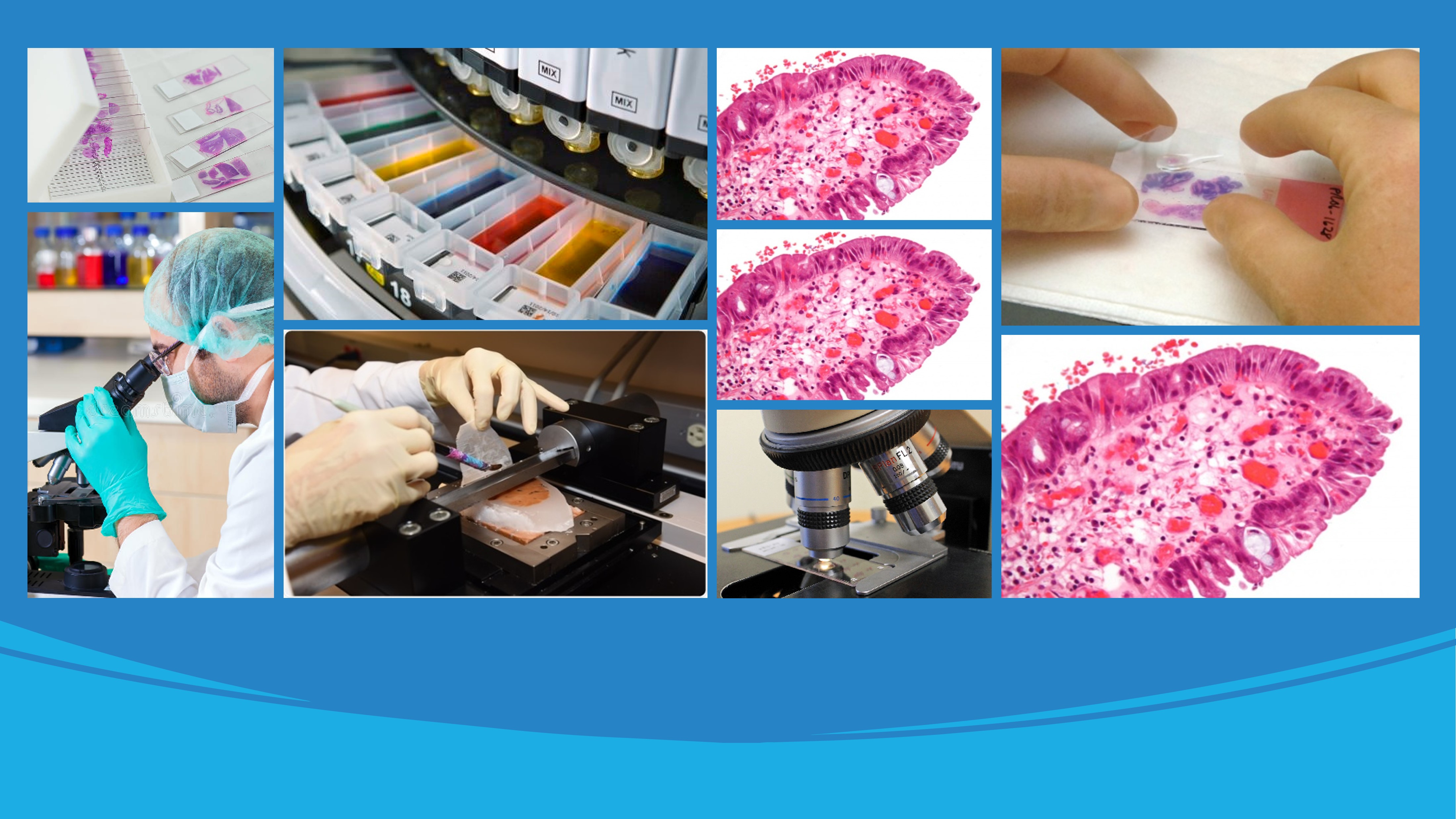
Rigorous clinical validation will require presenting the ADP database and diagnostic tools with a large sample of slides representing a range of normal and diseased states. St. Michael's Hospital (SMH) and University Health Network (UHN) will provide a total of 180,000 slides for the completion of this project in five years. The breakdown of the slides will be 130,000 slides from GI and 50,000 slides from Brain. The GI slides will include 22,000 normal anatomy and 108,000 disease slides across diverse organs, i.e. colon, small intestine, stomach, and esophagus. The diseased slides will contain diverse inflammatory and neoplastic diseases. All slides will be hematoxylin and eosin-stained slides of biopsy specimens retrieved from the archives of the Division of Pathology at the SMH. The Brain slides will include 8,000 normal and 42,000 disease slides across diverse regions of the central nervous system including cortical and cerebellar gray and white matter, brain stem, spinal cord and peripheral nerve.
Check it out
The challenge is to create a comprehensive ADP covering a broad spectrum of organs and tissues in a relative short period of time, a process that is manually intensive requiring experts (pathologists) time for annotation that is expensive and not readily available. We then plan to introduce a new methodology to incrementally collect, annotate and validate a comprehensive ADP database that can cover broad spectrum of GI and Neuro histology organs for supervised learning. The annotation workflow is empowered by HistoNet (trained on each data collection trial) to facilitate the validation process throughout a seamless interaction between our engineering and pathologists' teams. The WSI scans acquired with the Huron TissueScope brightfield scanner are divided into smaller image patches where a selective procedure is applied via HistoNet (a pre-trained CNN classifier) to predict the possible tissue types that exist in a patch, where each patch along its predicted labels is provided to an expert pathologist to review/edit the labels. Once a certain batch of image patches are revised, they will be augmented to the existing ADP and the HistoNet training will be further fine-tuned accordingly.
Check it out